Annalise.ai
AI clinical decision support for radiology, designed so a radiologist could trust it inside a thirty-second read.
00
problem
Radiology is overloaded and under-supplied, and a careless AI makes it worse: a model that flags too much adds noise a tired clinician has to clear. Annalise's model detected well across chest X-ray and CT brain; adoption turned on whether a radiologist would trust its output enough to act on it inside a thirty-second read. That trust layer was my job. I joined after the model had been training for a year and engineering had started. Before full Covid-19 lockdowns I fit clinic visits into the few weeks available, then ran all interviews, prototyping and usability testing with Australian and international clinics.
solution
Most AI products hide uncertainty to look confident. We showed the model's probability and the interval around it, so a clinician who could see where the model was unsure kept their own judgement in the loop. We patented that interface. Competitors showed a region of interest. We showed a per-finding pixel map with a confidence for every finding type. Clinicians cannot assess an AI they cannot see, and the field was under criticism for opacity.
Showing what the AI saw, and where it was unsure
Radiologists repeatedly told us that an AI which flags too much becomes another source of work. They needed to see the evidence behind a finding to judge whether it was credible. So each finding showed the model's probability and a pixel-level heatmap marking where on the image it sat.
The heatmap came from how the model already worked. It calculated a probability for every pixel as it assessed a scan, but the map was not being stored anywhere. Once we could see it existed, engineering had a clear case to keep it and bring it into the product. Before the application was built, I had real patient cases run through the early model and made a prototype from the findings and heatmaps it returned.
A radiologist could move from a finding straight to the relevant part of the image, see why the AI had raised it and decide whether to confirm or dismiss it. I also worked to cut what I called eye miles, the distance a radiologist's eyes travel across three or four monitors, and gave radiologists control over how the app behaved. This design for confidence and uncertainty was later granted an Australian patent.
Deciding what earns a radiologist's attention
The initial engineering approach ranked findings by model confidence, with the highest probability at the top. Our first reader study showed the problem. A rib fracture detected at 95% could sit above a suspected pneumothorax detected at 40%, and missing the pneumothorax is far more dangerous.
With the clinical team, I helped move the ranking to clinical consequence, so potentially life-threatening findings came first even when the model was less confident. We split the interface into Priority and Other findings. Separately from the app, we built integrations into the radiologist's worklist that moved urgent cases forward on the same logic. In testing, radiologists identified critical conditions earlier, and the teams had a shared rule for later prioritisation calls.
The version with no interface
Some hospitals used legacy PACS workstations that could only receive static DICOM secondary captures. On those, finding text had to be burned directly into the image, and every pixel used to explain a finding took space from the diagnostic image.
The same output had to work for a radiologist reading their hundredth case of the day and for a non-specialist in an emergency department seeing it for the first time. I defined the typography, contrast and layout across English, non-Latin scripts and different monitor resolutions. We also put structured finding data into custom DICOM tags, so the information could travel with the record and leave the image itself unchanged.
93% of clinicians still using it after two months
Annalise went on to process more than 2.1 million cases across 12,000 facilities worldwide. In post-rollout research, 93% of clinicians were still using the product after two months and 90% said it positively affected their reporting. Those figures measure the product. The model's diagnostic accuracy was the work of the clinical and data science teams.
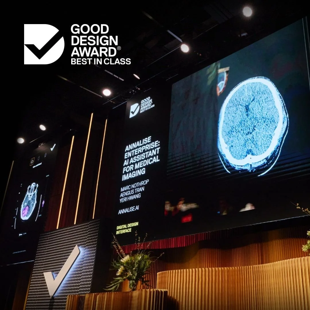
I built the design system that carried the same core interaction across four diagnostic modalities. The work won an IxDA Interaction Award (Best in Category, shortlisted alongside Google and Philips), Good Design Australia Best in Class and Good Design Korea.
What moved engineering, the founders and clinicians on costly or contested decisions was research and testing evidence showing precisely what the clinical user needed.
year
2020–2024
role
Founding Product Designer
what i made
User interviewing / Design, prototype & usability testing / Desktop application / Web & tablet app / Mobile X-ray embedded / Static rendered report / TGA/FDA reporting
01

02

03

04

05

see also

